
How Ultrasound Guidance Improves Achilles Tendon Injection Accuracy
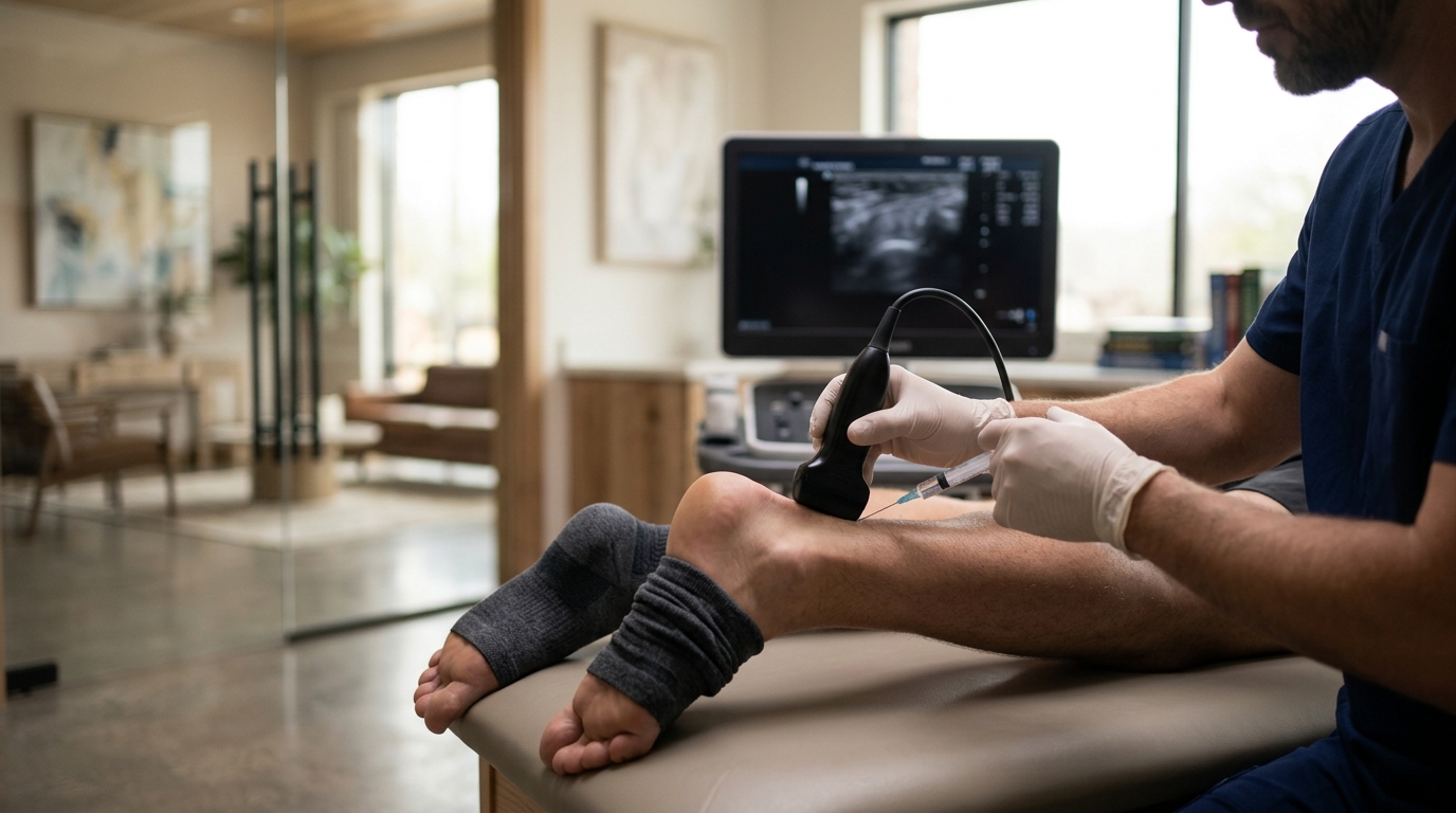
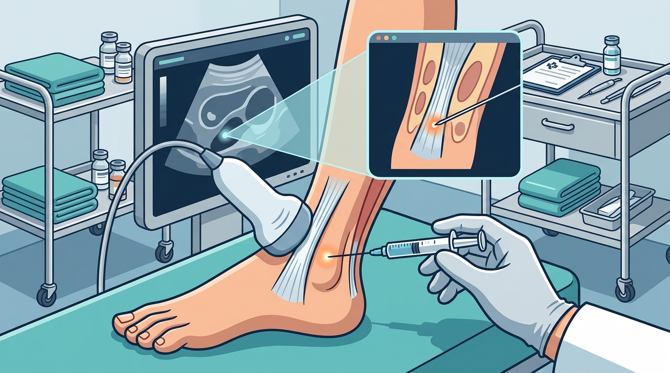
Ultrasound guidance improves Achilles tendon injection accuracy by allowing the clinician to visualize the needle tip in real time. This visualization ensures the medication is deposited directly into the affected tissue rather than surrounding structures. At Leroy Butler, DO PLLC, this precision is a standard part of our advanced ankle care protocol in Dallas and Frisco. By confirming needle placement, we reduce the risk of inadvertent injury and maximize the therapeutic effect of the injection.
The Anatomy of the Achilles Tendon
Blind injections, which rely on palpation alone, carry a higher risk of missing the target. The tendon is located superficially, but its relationship to the bone and surrounding soft tissue is not always obvious to the touch. Misplacement can lead to ineffective treatment or unnecessary pain. Visualizing the anatomy allows the surgeon to navigate these structures safely. For additional details, review the Notice of Privacy Practices.
Why Blind Injections Are Risky
Blind techniques lack the feedback loop that imaging provides. A clinician cannot see if the needle has penetrated the tendon or entered the bursa. This uncertainty can result in the medication being deposited in the wrong location. In some cases, the needle may inadvertently damage the tendon fibers. Ultrasound eliminates this guesswork by providing a live map of the injection site.
Real-Time Visualization and Needle Tracking
For Achilles tendinopathy, the goal is often to treat the degenerative changes within the tendon substance. This is known as an intratendinous injection. Ultrasound confirms that the needle tip is inside the tendon, not just in the surrounding paratenon. This distinction is vital for the success of the treatment. It ensures the medication reaches the cells responsible for the pain and inflammation.
The Role of High-Frequency Probes
High-frequency ultrasound probes are essential for imaging superficial structures like the Achilles tendon. These probes provide high-resolution images that can distinguish between healthy and degenerated tissue. They allow the clinician to assess the thickness and echotexture of the tendon. This assessment helps in planning the injection trajectory and depth.
Comparing Blind vs. Ultrasound-Guided Techniques
| Visualization | None | Real-time imaging |
| Needle Tracking | Palpation only | Visible on screen |
| Accuracy | Variable | High precision |
| Risk of Misplacement | Higher | Lower |
Improving Clinical Outcomes
Accurate placement directly correlates with better clinical outcomes. When medication is delivered to the precise site of pathology, the therapeutic effect is enhanced. Patients often report faster pain relief and improved function. This is because the treatment addresses the root cause rather than just the symptoms.
Studies have shown that image-guided injections can reduce the number of repeat procedures needed. By getting it right the first time, the overall treatment burden is lowered. This is particularly important for patients with chronic tendinopathy. It allows for a more efficient path to recovery. At Leroy Butler, DO PLLC, we prioritize these evidence-based techniques to ensure our patients receive the best possible care.
Patient Comfort and Safety
Ultrasound guidance also enhances patient comfort. The procedure is less invasive when the needle path is optimized. The clinician can choose a trajectory that avoids sensitive nerves and blood vessels. This reduces the risk of bruising and post-procedure pain. Patients often find the procedure less intimidating when they know it is being performed with visual precision.
The Role of Fellowship-Trained Expertise
Interpreting ultrasound images requires specialized training. A fellowship-trained orthopedic surgeon has the expertise to identify subtle changes in tendon structure. This training goes beyond basic medical education. It involves hundreds of hours of hands-on experience with musculoskeletal ultrasound. This expertise ensures that the images are interpreted correctly and the injection is performed safely.
Why Subspecialty Matters
General orthopedic surgeons may treat a wide range of conditions. However, subspecialists focus on specific areas of the body. This focus allows for deeper knowledge and more refined techniques. For Achilles tendon issues, this means a more nuanced approach to diagnosis and treatment. It ensures that the patient receives care that is calibrated to the specific demands of the foot and ankle.
Key Takeaways
- Ultrasound guidance allows for real-time visualization of the needle tip.
- It ensures medication is deposited directly into the affected tendon tissue.
- Blind injections carry a higher risk of misplacement and reduced efficacy.
- High-frequency probes provide the resolution needed for superficial structures.
- Accurate placement leads to faster pain relief and improved function.
- Fellowship-trained surgeons have the expertise to interpret ultrasound images accurately.
- Image-guided techniques can reduce the need for repeat procedures.
Frequently Asked Questions
Is ultrasound guidance painful?
How long does the procedure take?
The procedure typically takes 15 to 30 minutes. This includes the time needed to position the patient and perform the imaging. The actual injection is quick once the needle is in place.
Can ultrasound detect tendon tears?
Yes, ultrasound is highly effective at detecting partial and full-thickness tears. It can also assess the extent of degeneration and inflammation. This information helps in planning the appropriate treatment.
Is radiation involved in ultrasound?
No, ultrasound does not use ionizing radiation. It is a safe imaging modality that can be repeated as often as necessary. This makes it ideal for monitoring healing over time.
Who should perform ultrasound-guided injections?
The procedure should be performed by a clinician with specialized training in musculoskeletal ultrasound. Fellowship-trained orthopedic surgeons are well-suited for this task due to their expertise in anatomy and imaging.
How does this differ from MRI?
Ultrasound is real-time and allows for dynamic assessment. MRI provides a more detailed static image but cannot guide the needle in real time. Ultrasound is the preferred modality for guiding injections.
Conclusion
Ultrasound guidance is a critical tool in the treatment of Achilles tendinopathy. It enhances accuracy, safety, and clinical outcomes. By visualizing the needle in real time, clinicians can ensure that the medication reaches the target tissue. This precision is a hallmark of modern orthopedic care. At Leroy Butler, DO PLLC, we integrate this technology into our practice to provide the highest standard of care for our patients in Dallas and Frisco. If you are experiencing persistent heel pain, consider seeking a consultation with a fellowship-trained specialist who utilizes image-guided techniques.